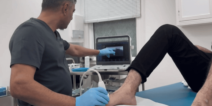
Ultrasound-Guided Cortisone Injections for Achilles Tendinopathy
Achilles tendinopathy is a common condition affecting the Achilles tendon, which connects the calf muscles to the heel bone. This condition is characterised by pain, stiffness, and reduced function in the tendon, often developing due to repetitive strain or overuse. While it is prevalent among athletes, particularly runners, it can also affect individuals who engage in activities that place excessive stress on the tendon.
Management options for Achilles tendinopathy vary depending on the severity and chronicity of the condition. Among the available treatment modalities, ultrasound-guided cortisone injections have gained attention as a potential intervention for managing tendon-related pain and inflammation.
This blog will provide a comprehensive overview of Achilles tendinopathy, including its anatomy, pathology, risk factors, symptoms, diagnosis, and various management approaches. It will also explore the role of ultrasound-guided cortisone injections in managing Achilles tendinopathy, detailing their mechanism of action and potential benefits.

Anatomy of the Achilles Tendon
The Achilles tendon is the strongest and largest tendon in the human body. It originates from the gastrocnemius and soleus muscles in the calf and inserts into the calcaneus (heel bone). This tendon plays a critical role in movements such as walking, running, and jumping, allowing for plantarflexion of the foot (pointing the toes downward).
The Achilles tendon has a limited blood supply, particularly in the midportion of the tendon, which makes it more susceptible to injury and delayed healing. The tendon is surrounded by a sheath called the paratenon, which facilitates smooth movement.
Pathology of Achilles Tendinopathy
Achilles tendinopathy is considered a degenerative condition rather than a purely inflammatory one. It is characterised by:
- Collagen disorganisation: Normal collagen fibres are aligned in a parallel manner, but in tendinopathy, they become disorganised and fragmented.
- Neovascularisation: The development of new blood vessels, often accompanied by nerve growth, which may contribute to pain.
- Increased cellular activity: Changes in tenocyte (tendon cell) activity can lead to increased production of matrix proteins and inflammatory mediators.
- Microtears and degeneration: Repetitive strain can lead to microscopic tears within the tendon, contributing to chronic pain and dysfunction.
Achilles tendinopathy is classified into two main types:
- Midportion Achilles Tendinopathy — Affects the main body of the tendon, typically 2–6 cm above the heel insertion.
- Insertional Achilles Tendinopathy — Occurs at the point where the tendon attaches to the heel bone, often associated with calcification or bony spurs.
Causes and Risk Factors
Several intrinsic and extrinsic factors contribute to the development of Achilles tendinopathy:
Intrinsic Factors:
- Age-related tendon degeneration
- Poor tendon blood supply
- Biomechanical issues (e.g., flat feet or high arches)
- Weak calf muscles or muscle imbalances
- Systemic conditions such as diabetes or rheumatoid arthritis
Extrinsic Factors:
- Sudden increase in activity level (e.g., increased running distance or intensity)
- Inadequate footwear with poor heel support
- Training errors (e.g., excessive hill running)
- Hard or uneven running surfaces
- Use of fluoroquinolone antibiotics, which may increase tendon vulnerability
Symptoms of Achilles Tendinopathy
Individuals with Achilles tendinopathy may experience the following symptoms:
- Gradual onset of pain at the back of the heel or along the tendon
- Stiffness and discomfort, particularly in the morning or after periods of rest
- Swelling or thickening of the tendon
- Tenderness when pressing on the affected area
- Pain that worsens with activity, especially running or jumping
- A creaking or crackling sensation when moving the tendon
Diagnosis of Achilles Tendinopathy
A clinical evaluation is essential for diagnosing Achilles tendinopathy. The diagnostic process typically includes:
- Medical History — Evaluating symptoms, activity levels, and risk factors.
- Physical Examination — Assessing for tenderness, swelling, and range of motion.
- Ultrasound Imaging — Providing detailed visualisation of tendon structure, detecting thickening, neovascularisation, and microtears.
Ultrasound imaging is particularly valuable in guiding cortisone injections to ensure precise delivery of the medication.
Management of Achilles Tendinopathy
Treatment strategies for Achilles tendinopathy typically involve a combination of conservative and interventional approaches.
Conservative Management
- Activity Modification — Reducing activities that exacerbate symptoms while maintaining general fitness.
- Eccentric Exercises — Strengthening the Achilles tendon through controlled lengthening exercises.
- Footwear and Orthotics — Wearing supportive shoes or heel lifts to reduce strain.
- Shockwave Therapy — A non-invasive therapy aimed at stimulating tendon healing.
Interventional Management
- Ultrasound-Guided Cortisone Injections — Used selectively for pain relief in cases of persistent discomfort.
Ultrasound-Guided Cortisone Injections for Achilles Tendinopathy
Cortisone injections are a type of corticosteroid treatment used to manage inflammation and pain in musculoskeletal conditions. When administered under ultrasound guidance, the injection can be precisely targeted to the affected area, improving accuracy and minimising potential complications.
Mechanism of Action
Cortisone injections work by:
- Reducing Inflammation — Corticosteroids inhibit inflammatory mediators, helping to alleviate swelling and pain.
- Suppressing Neovascularisation — The reduction in new blood vessel formation may help decrease pain associated with nerve growth.
- Modulating Cellular Activity — Corticosteroids can influence tendon cell activity, temporarily relieving symptoms.
Ultrasound guidance enhances the safety and effectiveness of the procedure by ensuring accurate placement of the injection.
Benefits of Ultrasound-Guided Cortisone Injections
- Precision — Ultrasound imaging ensures accurate placement of the medication.
- Real-Time Visualisation — Allows monitoring of needle positioning.
- Minimised Risk — Reduces the likelihood of inadvertent damage to surrounding structures.
Considerations and Precautions
While cortisone injections can provide temporary pain relief, their use in Achilles tendinopathy should be carefully considered. Repeated injections may be associated with an increased risk of tendon weakening or rupture. Therefore, they are typically reserved for cases where pain significantly impacts function and conservative treatments have not provided relief.
Conclusion
Achilles tendinopathy is a challenging condition that requires a comprehensive approach to management. While conservative treatments remain the first line of intervention, ultrasound-guided cortisone injections may be considered in cases of persistent pain. By precisely targeting the affected area, these injections can offer relief, allowing patients to engage in rehabilitation strategies effectively.
At Joint Injections, we specialise in ultrasound-guided injection therapies, ensuring precision and safety in treatment delivery. If you are experiencing Achilles tendon pain, a thorough assessment can help determine the most suitable management plan for your condition.
Comments
Post a Comment